Symptom Checker
Management
Toxic Plants A-Z
Pasture Grasses Comparison
Health
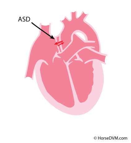
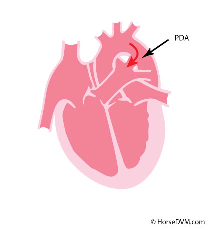
Diseases/Conditions A-Z
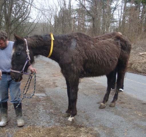
Horse Case Stories
Symptom Reference
Vaccines
Treatments
Drugs A-Z
Deworming
Nutrition
Equine Commercial Feeds A-Z
Equivalent Feed Search
Byproducts Comparison
Body Condition Scoring
Directory
Equine Veterinarians
Diagnostic Labs
Tools
Switch Animal
Poultry
Cow
Goat
Home
Symptoms
%
Diagnosis
While waiting for your veterinarian
Support
Therapies
Prevention
Scientific Research
Geographical Distribution
Risk Factors
Horse Case Stories