A keratoma is a benign tumor of the hoof that grows inside the horse's foot. It originates from the keratin-containing tissue, usually underneath the coronet, and between the hoof wall and third phalanx. As the size of the keratoma increases, it will exert pressure on the horse's sensitive laminae and surrounding structures in the hoof, causing pain and lameness.
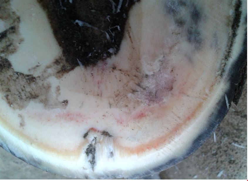
When keratomas reach the white line area at the toe of the foot, it will cause separation of the bond between the hoof wall and sole of the foot. If bacteria gets into the foot, it causes an abscess to develop, which look just like any other foot abscess. However, when horses have an underlying keratoma it will cause the abscess to keep recurring.
Clinical Signs
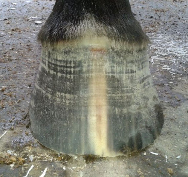
Horses with a keratoma often have a history of a recurring abscess in the same foot. They often show progressive lameness in the affected foot, graded at 3 to 4 out of 5. Some affected horses may have a visible deviation or bulge at the coronary band or abnormal hoof growth overlying the region containing the mass. The bulge will also be very painful when touched.
Diagnosis
Keratomas are diagnosed based on history (of recurring abscesses in the same foot), clinical signs, and radiographs of the foot. Radiographs will show the appearance of an indentation in the coffin bone inside the hoof, due to the loss of calcium in the bone.
Treatment
Treatment for a keratoma is surgical removal, either under general anesthesia or standing sedation. MRI-guided surgery can help reduce healing time by allowing for a more accurate determination of the tumor's location. Prolonged post-surgical care is required, since the hoof takes at least 10 to 12 months to grow back. Frequent bandage changes are required, with wound dressings applied regularly to control bleeding and infection. A special shoe is often applied to help stabilize the weakened hoof during the recovery period.
Once removed, keratomas don't usually grow back, and since they are a benign tumor, they don't spread.